Enlighten Knowledge note; The Lymphatic System.
The Lymphatic System.
The lymphatic system, which is closely associated with the cardiovascular system, has four main functions that contribute to homeostasis:
- Lymphatic capillaries absorb excess tissue fluid and return it to the bloodstream.
- In the small intestines, lymphatic capillaries called lacteals absorb fats in the form of lipoproteins and transport them to the bloodstream.
- The lymphatic system is responsible for the production, maintenance, and distribution of lymphocytes.
- The lymphatic system helps defend the body against pathogens.
Lymphatic Vessels
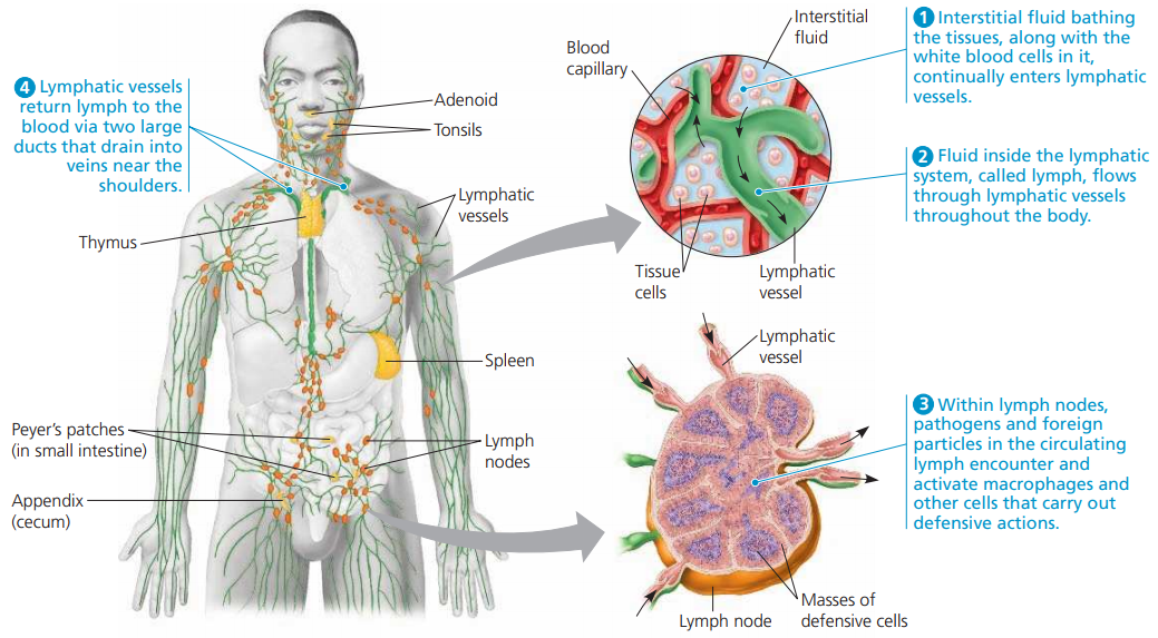
Lymphatic vessels form a one-way system that begins with lymphatic capillaries.
Most regions of the body are richly supplied with lymphatic capillaries tiny, closed-ended vessels.
Lymphatic capillaries take up excess tissue fluid.
The fluid inside lymphatic capillaries is called lymph.
In addition to water and fat molecules, lymph contains the same ions, nutrients, gases, and proteins that are present in tissue fluid.
It also contains defense molecules called antibodies, which are produced by lymphocytes.
The lymphatic capillaries join to form lymphatic vessels that merge before entering one of two ducts.
The thoracic duct or the right lymphatic duct.
The larger thoracic duct returns lymph to the left subclavian vein.
The right lymphatic duct returns lymph to the right subclavian vein.
The construction of the larger lymphatic vessels is similar to that of cardiovascular veins, including the presence of valves.
Skeletal muscle contraction forces lymph through lymphatic vessels, and then it is prevented from flowing backward by the one-way valves.
Lymphatic Organs
The organs of the lymphatic system consist of;
The primary lymphoid organs
- the bone marrow
- the thymus
The secondary lymphoid organs
- the lymph nodes,
- spleen,
- mucosa-associated lymphoid tissue, or MALT
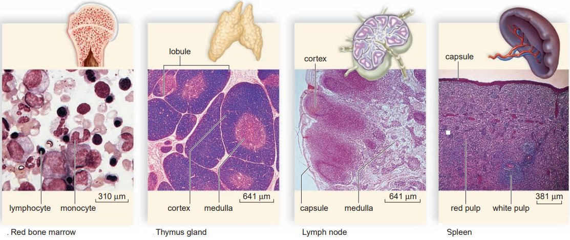
Red bone marrow:
Red bone marrow is a spongy, semisolid red tissue where stem cells divide and produce all the various types of blood cells, including lymphocytes these become mature B cells, a major type of lymphocyte, in the bone marrow.
In a child, most of the bones have red bone marrow
But in an adult, it is present only in the bones of the skull, the sternum (breastbone), the ribs, the clavicle (collarbone), the pelvic bones, the vertebral column, and the proximal heads of the femur and humerus.
The red bone marrow consists of a network of connective tissue fibers that supports the stem cells and their progeny.
They are packed around thin-walled sinuses filled with venous blood.
Differentiated blood cells enter the blood- stream at these sinuses.
The soft, bilobed thymus gland:
The soft, bilobed thymus gland is located in the thoracic cavity between the trachea and the sternum ventral to the heart.
Immature T cells, the other major type of lymphocyte, migrate from the bone marrow through the bloodstream to the thymus, where they mature.
The thymus also produces thymic hormones, such as thymosin, that are thought to aid in the maturation of T cells.
The thymus varies in size, but it is largest in children and shrinks as we get older. In the elderly, it is barely detectable. When well developed, it contains many lobules.
Lymph nodes:
They are small (about 1–25 mm in diameter), ovoid structures occurring along lymphatic vessels.
Lymph nodes are named for their location.
For example, inguinal lymph nodes are in the groin, and axillary lymph nodes are in the armpits.
Physicians often feel for the presence of swollen, tender lymph nodes in the neck as evidence that the body is fighting an infection.
- As lymph courses through the sinuses (open spaces) of the medulla.
- It is cleansed by macrophages, which engulf debris and pathogens.
- The many B and T cells also present help the immune system destroy pathogens.
Unfortunately, cancer cells sometimes enter lymphatic vessels and congregate in lymph nodes.
Therefore, when a person undergoes surgery for cancer, it is a routine procedure to remove some lymph nodes and examine them to determine whether the cancer has spread to other regions of the body.
The spleen:
The spleen, an oval organ with a dull purplish colour, is located in the upper left side of the abdominal cavity posterior to the stomach.
Most of the spleen is red pulp that filters and cleanses the blood.
Red pulp consists of blood vessels and sinuses, where macrophages remove old and defective blood cells.
The spleen also has white pulp that is inside the red pulp and consists of little lumps of lymphatic tissue where B and T cells congregate.
The spleen’s outer capsule is relatively thin, and an infection or a blow can cause the spleen to burst.
Although the spleen’s functions can be largely replaced by other organs, a person without a spleen is often slightly more susceptible to infections and may require antibiotic therapy indefinitely.
Patches of lymphatic tissue in the body include;
- The tonsils, located in the pharynx;
- Peyer patches, located in the intestinal wall.
- The vermiform appendix, attached to the cecum.
These structures encounter pathogens and antigens that enter the body by way of the mouth.
Join Enlighten Knowledge WhatsApp Channel.
Join Enlighten Knowledge Telegram platform.


Bravo