Mechanism of Sleep and Language and Spatial Recognition.
Mechanism of Sleep and Language and Spatial Recognition.
Arousal and Sleep.
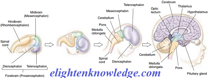
- The brain stem contains a diffuse collection of neurons referred to as the reticular formation.
- One part of this formation, the reticular activating system, controls consciousness and alertness.
- All of the sensory pathways feed into this system, which monitors the information coming into the brain and identifies important stimuli.
- When the reticular activating system has been stimulated to arousal, it increases the level of activity in many parts of the brain.
- Neural pathways from the reticular formation to the cortex and other brain regions are depressed by anesthetics and barbiturates.
- The reticular activating system controls both sleep and the waking state.
- It is easier to sleep in a dark room than in a light one because there are fewer visual stimuli to stimulate the reticular activating system.
- In addition, activity in this system is reduced by serotonin, a neurotransmitter we previously discussed.
- Serotonin causes the level of brain activity to fall, bringing on sleep.
Sleep is not the loss of consciousness.
- Rather, it is an active process whose multiple states can be revealed by recording the electrical activity of the brain in an electroencephalogram (EEG).
- In a relaxed but awake individual whose eyes are shut, the EEG consists primarily of large, slow waves that occur at a frequency of 8 to 13 hertz (cycles per second).
- These waves are referred to as alpha waves.
- In an alert subject whose eyes are open, the EEG waves are more rapid (beta waves are seen at frequencies of 13 to 30 hertz) and are more desynchronized because multiple sensory inputs are being received, processed, and translated into motor activities.
- Theta waves (4 to 7 hertz) and delta waves (0.5 to 4 hertz) are seen in various stages of sleep.
- The first change seen in the EEG with the onset of drowsiness is a slowing and reduction in the overall amplitude of the waves.
- This slow-wave sleep has several stages but is generally characterized by decreases in arousability, skeletal muscle tone, heart rate, blood pressure, and respiratory rate.
- During REM sleep (named for the rapid eye movements that occur during this stage), the EEG resembles that of a relaxed, awake individual, and the heart rate, blood pressure, and respiratory rate are all increased.
- Paradoxically, individuals in REM sleep are difficult to arouse and are more likely to awaken spontaneously.
- Dreaming occurs during REM sleep, and the rapid eye movements resemble the tracking movements made by the eyes when awake, suggesting that dreamers “watch” their dreams.
Language and Spatial Recognition.
- Although the two cerebral hemispheres seem structurally similar, they are responsible for different activities.
- The most thoroughly investigated example of this lateralization of function is language.
- The left hemisphere is the “dominant” hemisphere for language, that is, the hemisphere in which most neural processing related to language is performed but in all 90% of right-handed people and nearly two-thirds of left-handed people.
- There are two language areas in the dominant hemisphere.
Wernicke’s area;
- Located in the parietal lobe between the primary auditory and visual areas, is important for language comprehension and the formulation of thoughts into speech.
Broca’s area;
- Found near the part of the motor cortex controlling the face, is responsible for the generation of motor output needed for language communication.
- Damage to these brain areas can cause language disorders known as aphasias.
- For example, if Wernicke’s area is damaged, the person’s speech is rapid and fluid but lacks meaning; words are tossed together as in a “word salad.”
- While the dominant hemisphere for language is adept at sequential reasoning, like that needed to formulate a sentence, the nondominant hemisphere (the right hemisphere in most people) is adept at spatial reasoning, the type of reasoning needed to assemble a puzzle or draw a picture.
- It is also the hemisphere primarily involved in musical ability, a person with damage to Broca’s speech area in the left hemisphere may not be able to speak but may retain the ability to sing
- Damage to the nondominant hemisphere may lead to an inability to appreciate spatial relationships and may impair musical activities such as singing.
- Even more specifically, damage to the inferior temporal cortex in that hemisphere eliminates the capacity to recall faces.
- Reading, writing, and oral comprehension remain normal, and patients with this disability can still recognize acquaintances by their voices.
- The nondominant hemisphere is also important for the consolidation of memories of nonverbal experiences.
Join Enlighten Knowledge WhatsApp platform.
Join Enlighten Knowledge Telegram platform.
Follow our:
FACEBOOK PAGE.