Skin Cancers and Other Sun-Related Skin Growths
Skin Cancers and Other Sun-Related Skin Growths
Skin cancer
Skin cancer is caused by the damage to the DNA that we have been discussing.
The cells begin dividing unevenly and rapidly, but the genetic material in the DNA is damaged by the sun.
This rapid dividing of the cells may take the form of small tumors, which we know as skin cancer.
There are three major kinds of skin cancer;
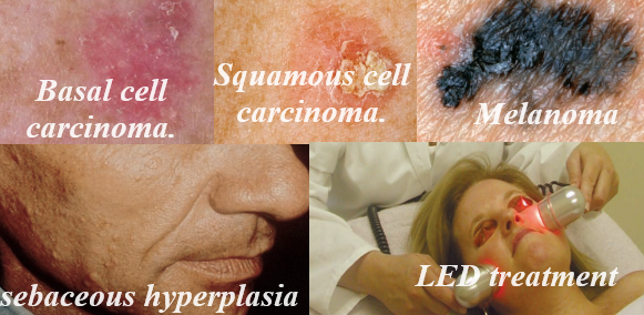
Basal cell carcinomas
- Basal cell carcinomas are the most common.
- They are almost always found in sun-exposed areas, typically on the face, hands, legs, back, and chest.
- They often look like small pearls or whitish, waxy nodules.
- Sometimes they have a small blood vessel running through them.
- They are not usually perfectly round like a milium.
- On older hyperpigmented skin, they may be hard to differentiate from milia.
- You must look carefully to tell the difference
- Aestheticians are not in the business of diagnosing skin cancer.
- However, you should know the signs of the major forms of skin cancer and be able to refer suspicious-looking lesions and areas to a dermatologist.
- Basal cell carcinomas are very often curable.
- In fact, 95 percent of these cancers are cured.
- They rarely spread to the internal parts of the body, but they can grow.
- If they are not treated, they can affect large areas of the skin.
- Basal cell carcinomas are sometimes removed surgically, sometimes frozen, and sometimes treated with laser or other means.
Squamous cell carcinomas
- Squamous cell carcinomas are the second most frequently diagnosed form of skin cancer.
- They may look like red or pink solid bumps on the skin.
- They may appear as open sores or ulcers that do not seem to heal.
- They may look crusty, or the client may notice a crusty area that bleeds easily.
- Many clients notice this when washing or cleansing the face.
- Again, these lesions appear in chronically sun-exposed areas.
- Treatment is similar to treatment for basal cell carcinoma, but squamous cell carcinoma may sometimes spread to other areas of the body.
- Mohs’ surgery is a specialized surgical technique, named after its developer, Frederic Mohs, M.D. In Mohs’ surgery, the dermatologist trims more and more tissue from the cancer lesion, carefully checking each sample of tissue for cancerous cells using a microscope, until all the cancerous tissue is removed.
Melanoma
- Melanoma is the least frequently seen form of the three major kinds of skin cancer and is also the deadliest.
- Melanoma is characterized by moles or mole-like lesions that are dark in colour.
- The Skin Cancer Foundation has coined an expression that is easy to remember when looking at suspicious lesions.
They call this the ABCDEs of Melanoma:
- A stands for asymmetric. The melanoma lesion usually grows to one side of the lesion and is uneven.
- B stands for border. The borders of melanomas are uneven and not smooth.
- C is for colour. Melanomas often have dark brown and black colours and are usually splotchy and not all one colour.
- D stands for diameter. Melanomas are usually at least the size of a pencil eraser or bigger. Regular moles are usually smaller than this.
- E stands for evolving. You must watch for changes and “evolving” moles. Changes may include darkening or variations in colour, moles that itch or hurt, and changes in the shape or growth of the mole.
Melanomas can be found on any area of the body but are most likely, again, to be found on areas that have had repeated sun exposure.
They are most typically found in individuals with a history of sunburn and light-skinned individuals.
Other sun-related growths
Actinic keratoses
- Actinic keratoses are rough areas of sun-damaged skin, indicated by dysplastic cell growth.
- Dysplastic means “abnormal growth.”
- Actinic keratoses are frequently found on the faces of individuals who have had chronic sun exposure.
- They are also prevalent in light-skinned individuals.
- They are rough patches of skin and can be crusty, scaly, and coarse to the touch.
- Sometimes they feel like small needles or splinters sticking out of the skin when they are touched with the fingers, and they are often noticed during applications of products or massage.
- They are frequently in the temple areas, forehead, and upper cheekbones.
- They frequently occur in small groups or patches.
- Because they are dysplastic cells, they can become cancerous and are often referred to as precancers.
Sebaceous hyperplasias
- Sebaceous hyperplasias are small, doughnut-shaped lesions that look like large, open comedones surrounded by a ridge of skin.
- Sebaceous hyperplasia actually means “overgrown oil glands.”
- These lesions are frequently seen on oily skin that has had repeated sun exposure, but they can appear on any skin, usually in someone age 25 and older.
- You will frequently find them on the forehead and temples, although they may appear anywhere on the face.
- Sebaceous hyperplasias are benign lesions that rarely cause any problems, except aesthetically.
- They are rarely removed surgically, because the scar from excision is usually worse than the appearance of the lesion.
- Sebaceous hyperplasia can be treated with electrodessication with an electric needle or sometimes, with cryosurgery.
- This helps flatten the lesion, but the patient may need to be retreated periodically because the oil glands are deep within the skin and can keep growing.
- Sebaceous hyperplasias are seen frequently by aestheticians, and clients are often concerned about them.
- Persons who have them also may have other symptoms of sun damage.
Seborrheic keratoses
- Seborrheic keratoses are large, flat, crusty-looking, brown, black, yellowish, grey or sometimes flesh-coloured lesions that are often found on the faces of older, sun-damaged clients.
- They are frequently found on the temples or cheekbones, although they can appear anywhere on the skin.
- They look almost like scab, and clients sometimes pick at them, which they obviously should not do.
- These growths are usually harmless but can occasionally turn into other more serious lesions, such as basal cell carcinomas.
- They are treated by the dermatologist, usually by curettage (a curette, a small, scoop-like instrument, is used to scrape the lesion off the skin).
- Seborrheic keratoses are more of a cosmetic nuisance than anything in most cases.
- Nevertheless, they should be referred to a dermatologist for treatment because they may indicate other areas or other forms of sun damage that may require treatment.
Solar lentigines
- Solar lentigines, often called solar freckles or “liver spots,” are skin areas containing “clumps” of hyperpigmentation caused by sun exposure.
- They can appear on any area but frequently appear on the face and hands.
- These can be treated by the aesthetician, and more severe cases can be treated by the dermatologist.
It is important to note that;
After a dermatologist treats skin cancer, the doctor may suggest that the client not have skin care treatments until the lesion is healed completely.
As a general rule, clients should not have facial treatment if a treated lesion still has sutures, or “stitches.”
Clients treated with fluorouracil should not have facial treatment until the therapy is completed unless approved by the dermatologist, who may occasionally refer such a client for help with side effects.
Make sure you have been properly trained by the doctor before treating these clients.
Other forms of growth treatment, such as electrosurgery or cryosurgery, generally do not cause any problem for treatment, except that facial treatment should not be performed until the lesion is healed or unless the doctor advises otherwise.
Avoid these areas completely, and simply treat the rest of the untreated areas of the face.
If you have any questions, contact the dermatologist.