Animal Tissues; Types and Their Functions.
Animal Tissues; Types and Their Functions.
Cells form a working animal body through their emergent properties, which arise from successive levels of structural and functional organization.
Cells are organized into tissues, groups of cells with a similar appearance and a common function.
Different types of tissues are further organized into functional units called organs. (The simplest animals, such as sponges, lack organs or even true tissues.)
Groups of organs that work together, providing an additional level of organization and coordination, make up an organ system.
Thus, for example, the skin is an organ of the integumentary system, which protects against infection and helps regulate body temperature.
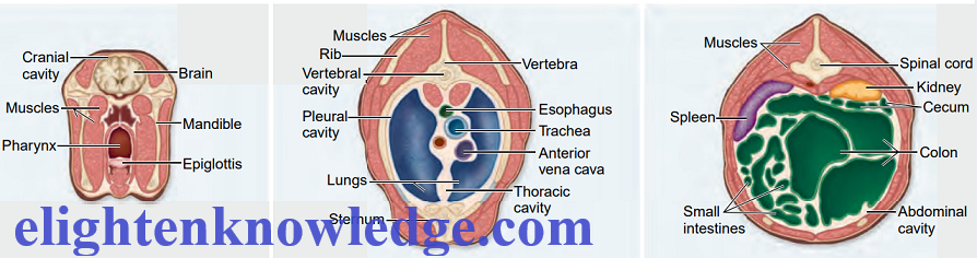
Animal Tissue.
A tissue is composed of specialized cells of the same type that perform a common function in the body.
The tissues of the human body can be categorized into four major types:
- Epithelial tissues.
- Connective tissues.
- Muscular tissues.
- Nervous tissue.
Epithelial Tissue:
Epithelial tissue, also called epithelium
It consists of tightly packed cells that form a continuous layer.
Epithelial tissue covers surfaces and lines body cavities.
Usually, it has a protective function, but it can also be modified to carry out
- secretion,
- absorption,
- excretion, and
- filtration.
Epithelial cells may be connected by three types of junctions composed of proteins.
- Tight junctions
- Adhesion junctions.
- Gap junctions.
Epithelia also form active interfaces with the environment but on the other side, they have a basement membrane.
The basement membrane is simply a thin layer of various types of proteins that anchors the epithelium to the extracellular matrix, which is often a type of connective tissue.
Types of epithelial tissues
-
- Simple Epithelia
- Complex Epithelia
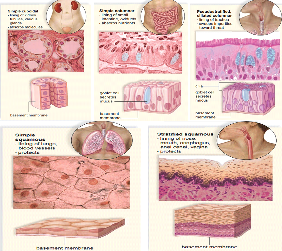
Simple Epithelia
Simple epithelia have only a single layer of cells and are classified according to cell type.
-
Simple squamous epithelia
Squamous epithelia are composed of flattened cells and are found lining blood vessels and the air sacs of the lungs.
-
Cuboidal epithelium
The cuboidal epithelium contains cube-shaped cells and is found lining the kidney tubules and various glands.
-
Columnar epithelium
This epithelium is found lining the digestive tract, where it efficiently absorbs nutrients from the small intestine because of minute cellular extensions called micro-villi.
The columnar epithelium has cells resembling rectangular pillars or columns, with nuclei usually located near the bottom of each cell.
The ciliated columnar epithelium is found lining the oviducts, where it propels the egg toward the uterus.
-
Pseudostratified columnar epithelium
When an epithelium is pseudostratified, it appears to be layered, but true layers do not exist because each cell touches the basement membrane.
The lining of the windpipe, or trachea, is pseudostratified ciliated columnar epithelium.
A secreted covering of mucus traps foreign particles, and the upward motion of the cilia carries the mucus to the back of the throat, where it may be either swallowed or expectorated.
Smoking can cause a change in mucus secretion and inhibit ciliary action, resulting in a chronic inflammatory condition called bronchitis.
-
Stratified Epithelia
Stratified epithelia have layers of cells piled one on top of the other.
Only the bottom layer touches the basement membrane.
The nose, mouth, oesophagus, anal canal, and vagina are all lined with stratified squamous epithelium.
Stratified cuboidal and stratified columnar epithelia also occur in the body.
-
Glandular Epithelia
When an epithelium secretes a product, it is said to be glandular.
A gland can be a single epithelial cell, as in the case of mucus-secreting goblet cells within the columnar epithelium lining the digestive tract, or a gland can contain many cells.
Glands that secrete their product into ducts are called exocrine glands.
Glands that have no ducts are appropriately known as endocrine glands.
-
Polarity of epithelia
All epithelia are polarized, meaning that they have two different sides.
The apical surface faces the lumen (cavity) or outside of the organ and is therefore exposed to fluid or air.
Specialized projections often cover this surface.
For example;
The apical surface of the epithelium lining the small intestine is covered with microvilli, projections that increase the surface area available for absorbing nutrients.
Opposite the apical surface of each epithelium is the basal surface.
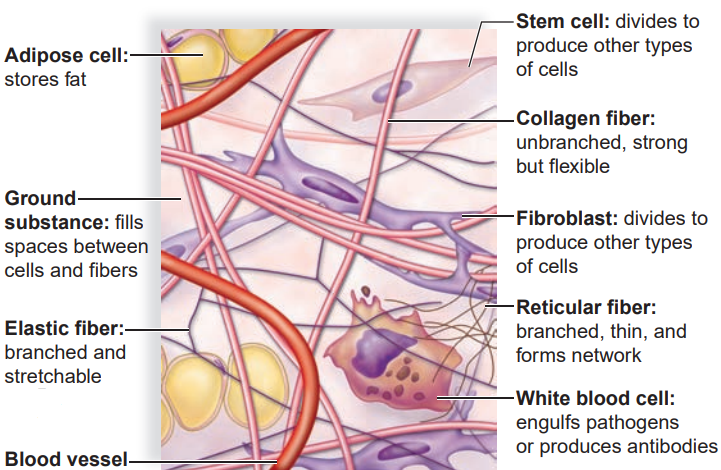
Connective Tissue
Connective tissue is the most abundant and widely distributed tissue in complex animals.
It is quite diverse in structure and function, but, even so, all types have three components:
- specialized cells
- ground substance
The ground substance is a noncellular material that separates the cells and varies in consistency from solid to semifluid to fluid.
- protein fibers
The fibers are of three possible types.
- White collagen fibers contain collagen, a protein that gives them flexibility and strength.
- Reticular fibers are very thin collagen fibers that are highly branched and form delicate supporting networks.
- Yellow elastic fibers contain elastin, a protein that is not as strong as collagen but is more elastic.
Connective tissue is classified into three categories:
- Fibrous Connective Tissue
- Supportive Connective Tissues
- Fluid Connective Tissues
Fibrous Connective Tissue
Fibrous connective tissue is of three types which are;
- Loose fibrous connective tissues
- Dense fibrous connective tissues
- Adipose tissue
Both loose fibrous and dense fibrous connective tissues have cells called fibroblasts that are located some distance from one another and are separated by a jellylike matrix containing white collagen fibers and yellow elastic fibers.
-
Loose fibrous connective tissue
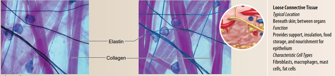
Loose fibrous connective tissue supports epithelium and also many internal organs.
Its presence in lungs, arteries, and the urinary bladder allows these organs to expand.
It forms a protective covering enclosing many internal organs, such as muscles, blood vessels, and nerves.
The most widespread connective tissue in the vertebrate body is loose connective tissue, which binds epithelia to underlying tissues and holds organs in place.
-
Dense fibrous connective tissue
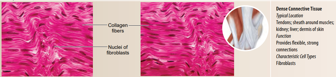
Dense fibrous connective tissue contains many collagen fibers that are packed together.
This type of tissue has more specific functions than does loose connective tissue.
For example, dense fibrous connective tissue is found in tendons, which connect muscles to bones, and in ligaments which connect bones to other bones at joints.
-
Adipose tissue
Commonly termed fat cells.
Adipose tissue serves as the body’s primary energy reservoir.
Each adipose cell contains a droplet of triglycerides within a storage vesicle.
When needed for energy, the adipose cell hydrolyzes its stored triglyceride and secretes fatty acids into the blood for oxidation by the cells of the muscles, liver, and other organs.
Adipose tissue also insulates the body, contributes to body contours, and provides cushioning.
In mammals, adipose tissue is found particularly beneath the skin, around the kidneys, and on the surface of the heart.
Adipose cells cannot divide and the number of adipose cells in an individual is fixed.
When a person gains weight, the cells become larger, and when weight is lost, the cells shrink.
In obese people, the individual cells called adipocytes may be up to five times larger than normal.
Most adipocytes are white, but in foetuses, infants, and children, they may be brown due to mitochondria and be good at heat production.
Supportive Connective Tissue
Cartilage

In cartilage, the cells lie in small chambers called lacunae, separated by a matrix that is solid yet flexible.
Unfortunately, because this tissue lacks a direct blood supply, it heals very slowly.
There are three types of cartilage, distinguished by the type of fiber in the matrix.
-
Hyaline cartilage
Hyaline cartilage is the most common type of cartilage
Contains only very fine collagen fibers.
The matrix has a white, translucent appearance.
Hyaline cartilage is found in the nose and at the ends of the long bones and the ribs, and it forms rings in the walls of respiratory passages.
The foetal skeleton also is made of this type of cartilage.
Later, the cartilaginous foetal skeleton is replaced by bone.
-
Elastic cartilage
Elastic cartilage has more elastic fibers than hyaline cartilage.
It is more flexible and is found in the framework of the outer ear.
-
Fibrocartilage
Fibrocartilage has a matrix containing strong collagen fibers.
Fibrocartilage is found in structures that withstand tension and pressure, such as the pads between the vertebrae in the backbone and the wedges in the knee joint.
Bone
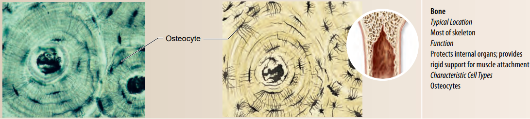
Bone is the most rigid of all the connective tissues.
It forms the skeleton, which supports the body, protects softer body structures such as the brain, and contributes to body movements.
Bone-forming cells called osteoblasts deposit a matrix of collagen.
Calcium, magnesium, and phosphate ions combine into a hard mineral within the matrix.
The microscopic structure of hard mammalian bone consists of repeating units called osteons.
Each osteon has concentric layers of the mineralized matrix, which are deposited around a central canal containing blood vessels and nerves.
Fluid Connective Tissues
-
Blood
Blood has a liquid extracellular matrix called plasma.
The consists of water, salts, and dissolved proteins.
Suspended in plasma are;
- Erythrocytes (red blood cells) = carry oxygen
- Leukocytes (white blood cells) = function in defence
- Cell fragments called platelets = (aid in blood clotting.)
Why is blood classified as a connective tissue?
We classify blood as a connective tissue because it contains abundant extracellular material, the fluid plasma.
The cells of blood are erythrocytes, or red blood cells, and leukocytes, or white blood cells.
Blood also contains platelets, or thrombocytes, which are fragments of a type of bone marrow cell.
Muscle Tissue
Muscles are the motors of the vertebrate body.
The characteristic that makes muscle cells unique is the relative abundance and organization of actin and myosin filaments within them.
Although these filaments form a fine network in all eukaryotic cells, where they contribute to the movement of materials within the cell, they are far more abundant and organized in muscle cells, which are specialized for contraction.
Vertebrates possess three kinds of muscle:
- Smooth muscle.
- Skeletal muscle.
- Cardiac muscle.
Skeletal and cardiac muscles are also known as striated muscles because their cells appear to have transverse stripes when viewed in longitudinal sections under the microscope.
The contraction of each skeletal muscle is under voluntary control, whereas the contraction of cardiac and smooth muscles is generally involuntary.
-
Smooth muscle.
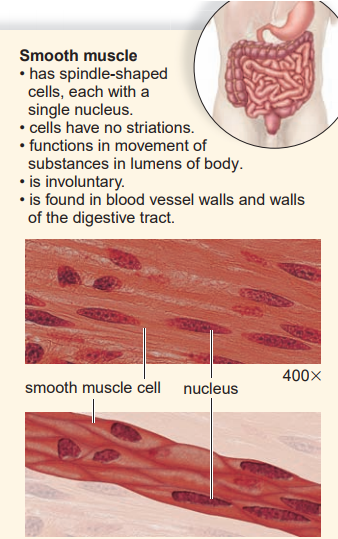
Smooth muscle was the earliest form of muscle to evolve.
It is found throughout most of the animal kingdom.
In vertebrates, smooth muscle occurs in the organs of the internal environment, or viscera, and is also called visceral muscle.
Smooth muscle tissue is arranged into sheets of long, spindle-shaped cells, each cell containing a single nucleus.
In some tissues, the cells contract only when a nerve stimulates them and then all of the cells in the sheet contract as a unit.
Some vertebrates, muscles of this type line the walls of many blood vessels and make up the iris of the eye, which contracts in bright light.
In other smooth muscle tissues, such as those in the wall of the digestive tract, the muscle cells themselves may spontaneously initiate electrical impulses, leading to a slow, steady contraction of the tissue.
Here nerves regulate, rather than cause, the activity.
-
Skeletal muscle
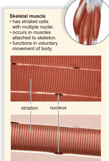
Skeletal muscles are usually attached to bones by tendons, so that their contraction causes the bones to move at their joints.
A skeletal muscle is made up of numerous, very long muscle cells called muscle fibers, which have multiple nuclei.
The fibers lie parallel to each other within the muscle and are connected to the tendons on the ends of the muscle.
Each skeletal muscle fiber is stimulated to contract by a motor neuron.
The nervous system controls the overall strength of a skeletal muscle contraction by controlling the number of motor neurons that fire, and therefore the number of muscle fibers stimulated to contract.
Each muscle fiber contracts by means of substructures called myofibrils containing highly ordered arrays of actin and myosin myofilaments.
These filaments give the muscle fiber its striated appearance.
Skeletal muscle fibers are produced during development by the fusion of several cells, end to end.
This embryological development explains why a mature muscle fiber contains many nuclei.
-
Cardiac muscle
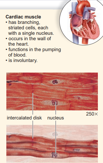
Cardiac muscle makes up the walls of the heart.
Its contraction pumps blood and accounts for the heartbeat.
Cardiac muscle combines features of both smooth muscle and skeletal muscle.
Like skeletal muscle, it has striations, but the contraction of the heart is involuntary for the most part.
Cardiac muscle cells also differ from skeletal muscle cells in that they have a single, centrally placed nucleus.
The cells are branched and seemingly fused one with the other, and the heart appears to be composed of one large interconnecting mass of muscle cells.
Actually, cardiac muscle cells are separate and individual, but they are bound end to end at intercalated disks, areas where folded plasma membranes between two cells contain adhesion junctions and gap junctions.
Nervous tissue.
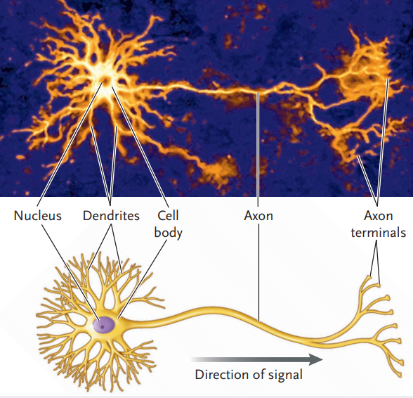
Nervous tissue contains nerve cells called neurons.
An average person has about 1 trillion neurons.
A neuron is a specialized cell that has three parts: dendrites, a cell body, and an axon.
A dendrite is a process that conducts signals toward the cell body.
The cell body contains the major concentration of the cytoplasm and the nucleus of the neuron.
An axon is a process that typically conducts nerve impulses away from the cell body.
Long axons are covered by myelin, a white, fatty substance.
The term fiber1 is used here to refer to an axon along with its myelin sheath if it has one.
Outside the brain and spinal cord, fibers bound by connective tissue form nerves.
The nervous system has just three functions:
- sensory input,
- integration of data, and
- motor output.
Nerves conduct impulses from sensory receptors to the spinal cord and the brain, where integration occurs. The phenomenon called sensation occurs only in the brain, however.
Nerves also conduct nerve impulses away from the spinal cord and brain to the muscles and glands, causing them to contract and secrete, respectively.
In this way, a coordinated response to the stimulus is achieved.
-
Neuroglia
Neuroglia are cells that outnumber neurons as much as 50 to 1.
It takes up more than half the volume of the brain.
Although the primary function of neuroglia is to support and nourish neurons, research is currently being conducted to determine how much they directly contribute to brain function.
Various types of neuroglia are found in the brain.
Microglia, astrocytes, and oligodendrocytes.
Microglia, in addition to supporting neurons, also engulf bacterial and cellular debris.
Astrocytes provide nutrients to neurons and produce a hormone known as glia-derived growth factor, which someday might be used as a cure for Parkinson disease and other diseases caused by neuron degeneration.
Oligodendrocytes form myelin.
Neuroglia do not have a long process, but even so, researchers are now beginning to gather evidence that they do communicate among themselves and with neurons.
Mature neurons have little capacity for cell division and seldom form tumors.
The majority of brain tumors in adults involve actively dividing neuroglia cells.
Most brain tumors have to be treated with surgery or radiation therapy because of a blood-brain barrier.
Join Enlighten Knowledge WhatsApp platform.
Join Enlighten Knowledge Telegram platform.
Follow our:
FACEBOOK PAGE.


Great and very educative. Keep up the good work
Thank you